Thursday, October 27, 2011

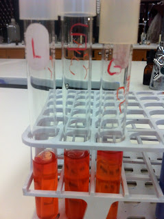
Its a pretty pink color :)

Today in lab we needed to check our plates we streaked before the field trip. The Mannitol Salt agar plate showed colonies that were yellow in color. This means that our bacteria creates acid as an end product. The fact that our bacteria grew means that is uses salt or is a halophile.


Then we checked our MacConkey plate. This plate only allows gram negative bacteria to grow. If the bacteria would have been red then it would be a coliform bacteria. Ours was not colored so it is a non-coliform.


The last plate we checked was the EMB or Eosin Metylene Blue agar plate. This plate allows only gram negative bacteria to grow. And if the bacteria was a pink colored that means it is lactose-fermented. This means it could be Enterobacter aerogenes.


We didn't do any other experiments because we have our Fall Break coming up this weekend and we won't be in lab to check results.


Tuesday, October 25, 2011

Field Trip!

We didn't have an actual lab today because we went to the water treatment plant for Steubenville water. We were able to go inside each building, see the pumps, see where the water is held at each stage of treatment, and got tons of information on how they do it, what they put in the water and where all it goes.

Before we left for the field trip we needed to inoculate an EMB plate, a Mannitol Salt Agar plate and a MacConkey's plate.


After we got back from the treatment plant, the last thing we needed to do was check our litmus milk test. The top was liquid while the bottom stayed solid.


Thursday, October 20, 2011

7 Test Tubes & 1 Positive Reaction

On Wednesday we needed to go into lab and document the reactions after 24 hours. There wasn't much change from Tuesday.
        lactose                                                         sucrose
       glucose                                                       comparison of all 3 sugars
       litmus milk                                                 MR-VP before adding reagents
      gelatin                                                          triple sugar iron

Today in lab we checked the 48 hour reactions and added reagents to the MR-VP experiment.
The three different sugar tests, lactose, sucrose and glucose, were negative meaning that our bacteria doesn't use carbohydrates to grow. The TSI test also showed the same result of negative.
The litmus milk test still showed no change. The gelatin test showed a small growth and a liquid portion at the top of the tube. This means that our bacteria is able to hydrolyze gelatin. For the MR-VP test we needed to add reagents "A" and "B" to 1mL and methyl red to the rest of the test tube. This made the test show a negative result.
      positive gelatin                                             "A"
     MR-VP with "A" & "B"                            Swirling for 5 minutes.....
   MR-VP with methyl red

Tuesday, October 18, 2011

Bubbles!

Today in lab we started the day looking at our results from last Thursday. Then Dr. Pathakamuri showed us another way to test for the presence of oxygen. This used hydrogen peroxide poured on our samples. The oxygen will bubble much like it does on human skin.
For the rest of lab we performed many different experiments. These included MR-VP (Methyl Red & Voges-Proskauer) to see if our unknown bacteria has the ability to ferment glucose. Each part of the experiment (MR and VP) have different reagents that need to be added after incubation. Another experiment was the Gelatin Hydrolysis where we stabbed a gelatin deep with an inoculating needle. The third experiment was the Triple Sugar Iron Agar test where we needed to stab the TSI tube with the needle 3/4 through the butt and make a zigzag pattern up the rest of the slant. The fourth test was the Litmus Milk Reaction where we inoculated a tube of litmus milk (that was a cool purple color) with our unknown. The final test was the Fermentation of Carbohydrates where we inoculated three test tubes, one containing a sucrose, glucose and lactose medium.

Sunday, October 16, 2011

Milk was a baaaad choice..

Last Friday we had to come in to check on the nutrient requirement experiments for our unknown bacteria.

This is what the starch agar plate looked like after we added the Gram's iodine. There is no halo around the growth which means that the experiment is negative for amylase production. There was no blue/black precipitate which would mean that there was starch present but it was negative for amylase production. Teresa and Lisa's half shows a positive result.
The skim milk agar plate showed a small halo around the growth which means it is positive for casein protease production. There could also have been an opaque white area which would mean there was casein present but it was negative for casein protease production.
The blue tributyrin plate showed a halo around the streak but it looks the opposite of the halo which would show a complete hydrolysis of triglycerides and a positive reaction for lipase production. Our unknown was negative. The other options could be that there could be a dark blue precipitate which would mean there was a partial hydrolysis of triglycerides and that it is positive for lipase production. Also the agar could be pale blue agar and no halo that would mean there was tributyrin present and it was negative for lipase production.

Thursday, October 13, 2011

What's For Dinner?

The answer is starch, skim milk, and fat but we'll get to that later.
Today in lab we looked at our test tube and plate from Tuesday to see what amount of oxygen our unknown needs to grow. From the test tube we were able to tell that it seemed to be obligate aerobic because all the grow was at the top of the liquid.
It looked like a jellyfish to me (Courtney). But when we looked at the bottom of the test tube there was a very small amount of growth and when we looked at the plate there was one single colony of growth. This shows that our bacteria is actually facultative but only slightly.
We also compared our unknown to other groups and with Theresa and Lisa who shared the plate with us. Their bacteria was truly facultative.

The next couple experiments we did were also grouped with Lisa and Theresa to save plates and agar. We needed to see which nutrients our bacteria uses to grow. The first test was to see the use of starch. For this we used a starch agar plate, which was a transparent color, with our bacteria on one side and their's on the other. The second was the same set up but on a skim milk agar plate that looked slightly yellow-orange and the third was on a tributyrin plate that was a cool blue color. The starch plate will show starch hydrolysis, the skim milk will show casein hydrolysis, and the tributyrin will show triglyceride or fat hydrolysis.


Tuesday, October 11, 2011

Micro-Aero .... Small Air ??

Today in lab we learned how to freeze bacteria to keep it to study or make vaccines later on. We needed to use 500 microliters of solution that was made up of 30% glycerol (150 microliters) and 70% broth (350 mircoliters). We put the glycerol in a microcentrifuge or cryovial tube. Then we added the broth culture. The tube was mixed and placed in a cyrostorage box that was placed in the freezer that was -70 degrees Celsius.
Then we did two different experiments to check if our bacteria needs oxygen or carbon dioxide to grow. First we inoculated a thioglycollate broth with our unknown. We already did this accidentally with the bacteria from the dollar bill the week before.
Our dollar bill bacteria is microaerophilic (which means that it likes small amounts of oxygen).
Then we needed to check for the use of growth with carbon dioxide. We got a petri dish and shared it with Theresa and Lisa (to use less plates and take up less space). The plate was split in half and we each streaked our unknown bacteria and put the plate in a GasPak anaerobic system. This tank decreases oxygen and increases carbon dioxide.


After all the groups got their plates together Dr. Pathakamuri showed us how to operate the system and I happened to record it...


Thursday, October 6, 2011

Stain gets on everything..

Today in lab we did the endospore stain and acid-fast stain. Both stains required to be performed over a boiling beaker of water on a heating plate. We fixed two smears and placed them on top of the beaker to heat. We placed bibulous paper over the slide and covered it with the correct stains for each procedure. Because it was heated we needed to make sure the slide did not dry. This used a lot of stain.


The endospore stain showed that our unknown does have endospores because the spores took up the green stain.
Our unknown didn't take up the acid-fast stain because our bacteria's cell walls don't contain the mycolic acid that is able to hold the red stain. The acid-alcohol washed away the red stain and allowed the blue stain to hold.
This lab got a little messy and we ended up having a little fun while everyone else finished their stains. (We were lucky enough to get to a heating plate before everyone else.)


Tuesday, October 4, 2011

Wait..What's Inoculating?

Today our lab was cut short because of the 11:00 classes being canceled. The only thing we needed to do was inoculate nutrient broth with our environmental sample off the dollar bill. This took us maybe about 2 minutes tops.
As for the title, when Dr. Pathakamuri was telling us that we just needed to inoculate there were more than a few people ask what inoculating was.. Not a good sign at this time in the semester.
To make up for missing the lab we also need to go see the new movie Contagion. Seems simple enough..

Monday, October 3, 2011

It Stained Our Fingerprints!

Today, September 27th, after our microbiology test we went into lab to do a capsule stain. To do this stain we placed a drop of nigrosin onto a slide. We then used the inoculating loop to place bacteria into the nigrosin drop. Using a clean slide we spread the nigrosin down the slide. We then let it air dry. Once the nigrosin was air dry we spread crystal violet stain over the nigrosin and waited for the stain to dry. Once the stain was dry we rinsed the excess off with water. We then blotted off the excess water and examined our new slides. Because of the capsule stain, we came to the conclusion that neither of our samples, the unknown and the dollar bill, had capsules. This is known because there was not a unstained circle around the dark bacteria.

before

during

after
As for the title, we apparently didn't clean the slide well enough and the nigrosin caught our fingerprints.